A Quantitative Proteomic Analysis of In Vitro Assembled Chromatin
25-Jan-2016
Molecular & Cellular Proteomics, vol. 15, no. 3, 945-959, doi: 10.1074/mcp.M115.053553
Molecular & Cellular Proteomics, online article
The structure of chromatin is critical for many aspects of cellular physiology and is considered to be the primary medium to store epigenetic information. It is defined by the histone molecules that constitute the nucleosome, the positioning of the nucleosomes along the DNA and the non-histone proteins that associate with it. These factors help
to establish and maintain a largely DNA sequence-independent but surprisingly stable structure. Chromatin is extensively disassembled and reassembled during DNA replication, repair, recombination or transcription in order to allow the necessary factors to gain access to their substrate. Despite such constant interference with chromatin
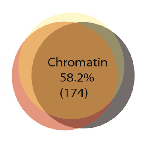
structure, the epigenetic information is generally well maintained. Surprisingly, the mechanisms that coordinate chromatin assembly and ensure proper assembly are not particularly well understood. Here, we use label free quantitative mass spectrometry to describe the kinetics of in vitro assembled chromatin supported by an embryo extract prepared from preblastoderm Drosophila melanogaster embryos. The use of a data independent acquisition method for proteome wide quantitation allows a time resolved comparison of in vitro chromatin assembly. A comparison of our in vitro data with proteomic studies of replicative chromatin assembly in vivo reveals an extensive overlap showing that the in vitro system can be used for investigating the kinetics of chromatin assembly in a proteome wide manner.


