Release pathways of interferon α2a molecules from lipid twin screw extrudates revealed by single molecule fluorescence microscopy
10-Sep-2012
Journal of Controlled Release, 2012, doi:10.1016/j.jconrel.2012.07.014, Volume 162, Issue 2, Pages 295–302 published on 10.09.2012
Journal of Controlled Release, online article
Journal of Controlled Release, online article
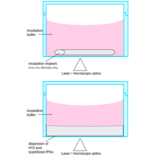
The pathways of interferon α2a release from a triglyceride based implant system were studied by single molecule fluorescence microscopy. The protein was labeled with a stable fluorescent dye ATTO647N, freeze-dried and embedded into the lipid matrix via twin-screw extrusion. The implant system consisted of a pore-forming agent (water soluble PEG 6000) and two types of triglycerides with different melting ranges which allowed the production of the implants at moderate temperatures and without the use of organic solvents. Single molecule microscopy and single particle tracking of labeled proteins contained in these implants revealed that two populations of diffusing proteins were present. Moreover, proteins were not only released via water-filled pores (created by dissolution of the pore-former), but surprisingly also through diffusion in a phase of molten lipid. Diffusion coefficients of IFNα 2a derived by tracking of individual protein molecules within the implant system were similar to diffusion coefficients obtained from control measurements in pure molten lipid and highly concentrated solutions of PEG 6000. In conclusion, tracking of individual protein molecules was successfully used to elucidate the release pathways of proteins from a relevant lipid based implant system.


