High contrast tumor imaging with radio-labeled antibody Fab fragments tailored for optimized pharmacokinetics via PASylation
21-Nov-2014
mAbs, Volume 7, Issue 1, pages 96-109, DOI:10.4161/19420862.2014.985522
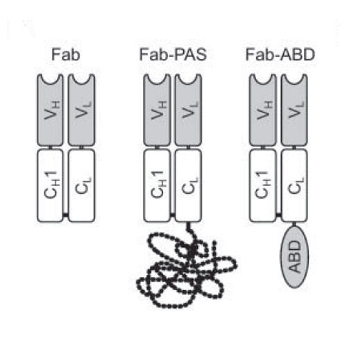
Although antigen-binding fragments (Fabs) of antibodies constitute established tracers for in vivo radiodiagnostics, their functionality is hampered by a very short circulation half-life. PASylation, the genetic fusion with a long, conformationally disordered amino acid chain comprising Pro, Ala and Ser, provides a convenient way to expand protein size and, consequently, retard renal filtration. Humanized αHER2 and αCD20 Fabs were systematically fused with 100 to 600 PAS residues and produced in E. coli. Cytofluorimetric titration analysis on tumor cell lines confirmed that antigen-binding activities of the parental antibodies were retained. The radio-iodinated PASylated Fabs were studied by positron emission tomography (PET) imaging and biodistribution analysis in mouse tumor xenograft models. While the unmodified αHER2 and αCD20 Fabs showed weak tumor uptake (0.8% and 0.2% ID/g, respectively; 24 h p.i.) tumor-associated radioactivity was boosted with increasing PAS length (up to 9 and 26-fold, respectively), approaching an optimum for Fab-PAS400. Remarkably, 6- and 5-fold higher tumor-to-blood ratios compared with the unmodified Fabs were measured in the biodistribution analysis (48 h p.i.) for αHER2 Fab-PAS100 and Fab-PAS200, respectively. These findings were confirmed by PET studies, showing high imaging contrast in line with tumor-to-blood ratios of 12.2 and 5.7 (24 h p.i.) for αHER2 Fab-PAS100 and Fab-PAS200. Even stronger tumor signals were obtained with the corresponding αCD20 Fabs, both in PET imaging and biodistribution analysis, with an uptake of 2.8% ID/g for Fab-PAS100vs. 0.24% ID/g for the unmodified Fab. Hence, by engineering Fabs via PASylation, plasma half-life can be tailored to significantly improve tracer uptake and tumor contrast, thus optimally matching reagent/target interactions


