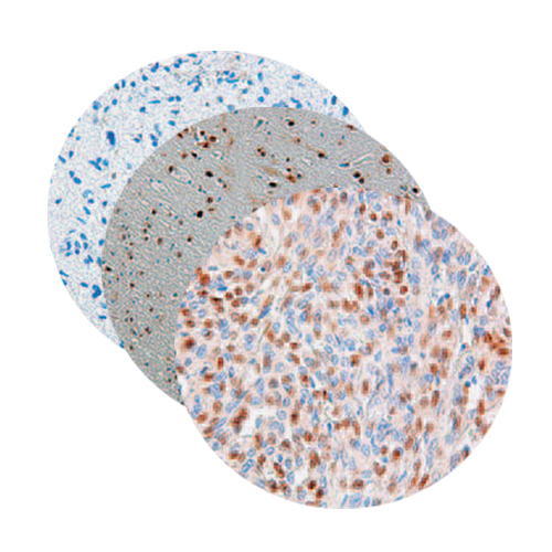
Low values of 5-hydroxymethylcytosine (5hmC), the ‘‘sixth base,’’ are associated with anaplasia in human brain tumors
10-Jan-2012
Int. J. Cancer, 2012, DOI: 10.1002/ijc.27429, 131, 1577–1590 published on 10.01.2012
Int. J. Cancer, online article
5-Methylcytosine (5 mC) in genomic DNA has important epigenetic functions in embryonic development and tumor biology. 5-Hydroxymethylcytosine (5 hmC) is generated from 5 mC by the action of the TET (Ten-Eleven-Translocation) enzymes and may be an intermediate to further oxidation and finally demethylation of 5 mC. We have used immunohistochemistry (IHC) and isotope-based liquid chromatography mass spectrometry (LC-MS) to investigate the presence and distribution of 5 hmC in human brain and brain tumors. In the normal adult brain, IHC identified 61.5% 5 hmC positive cells in the cortex and 32.4% 5 hmC in white matter (WM) areas. In tumors, positive staining of cells ranged from 1.1% in glioblastomas (GBMs) (WHO Grade IV) to 8.9% in Grade I gliomas (pilocytic astrocytomas). In the normal adult human brain, LC-MS also showed highest values in cortical areas (1.17% 5 hmC/dG [deoxyguanosine]), in the cerebral WM we measured around 0.70% 5 hmC/dG. levels were related to tumor differentiation, ranging from lowest values of 0.078% 5 hmC/dG in GBMs (WHO Grade IV) to 0.24% 5 hmC/dG in WHO Grade II diffuse astrocytomas. 5 hmC measurements were unrelated to 5 mC values. We find that the number of 5 hmC positive cells and the amount of 5 hmC/dG in the genome that has been proposed to be related to pluripotency and lineage commitment in embryonic stem cells is also associated with brain tumor differentiation and anaplasia.


